
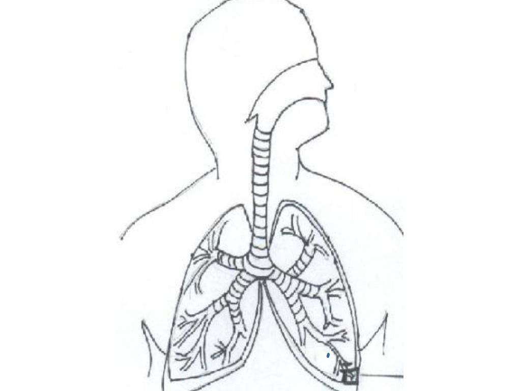
The Respiratory System Anatomy Sketch
This article will discuss the development and anatomy of the bronchopulmonary segments. It will also review the anatomy of the lungs, and discuss the function of the organs as well. Further clinical discussion involving disorders of the lung, as well as clinical investigation of pulmonary disorders will also be included. Key facts.
Human Lungs Outline Body Pages Coloring Colouring Printable Drawing Diagram Clip Lung Heart Kids
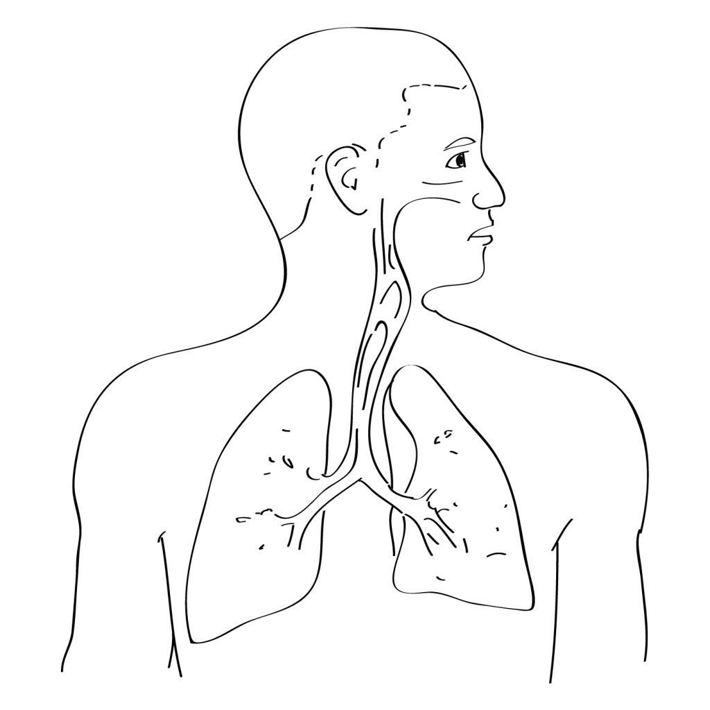
Answer link In the rib cage, around the heart They are in the central chest area
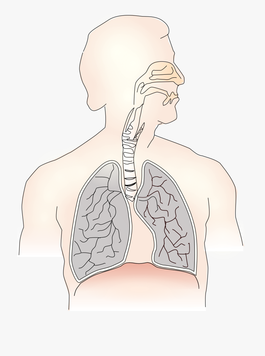
Respiratory system diagram unlabeled
Pulmonary Ventilation. Pulmonary ventilation is the act of breathing, which can be described as the movement of air into and out of the lungs. When you take a deep breath, notice the expansion of your rib cage. Contraction of the diaphragm and external intercostal muscles increases the volume in the chest cavity, which in turn lowers the pressure and draws air into the lungs for inspiration.
Where are the lungs located? Socratic
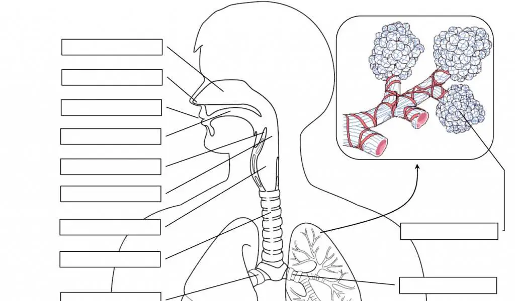
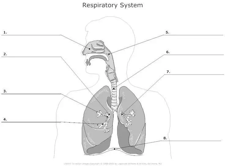
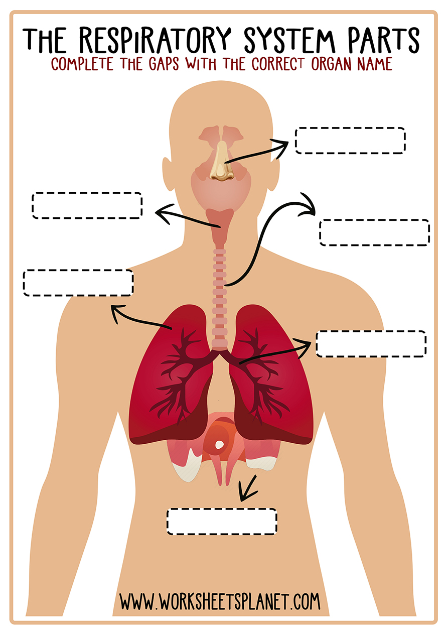
Printable Blackline Diagram of The Respiratory System Test yourself: fill in the blanks: Experiment: How do Your Lungs Work? Here is a simple project to show how your lungs work and how breathing happens. This lesson activity goes well with a study of anatomy, life science, and/or biology. https://www.lessontutor.com/km1/
Blank ipicture of basic anatomy of breathing garetkind
The lungs are a major organ that is part of the respiratory system, taking in fresh air and getting rid of old, stale air. This mechanism of breathing also helps to allow you to talk. By taking in fresh air, the lungs are able to help oxygenate blood to be carried around your body. This is done by inhaling the air and bringing it in toward the.
Download Unlabeled Lung Diagram Images Diagram Printabel
The Nasal Cavity. The nasal epithelium (Figure 20.6.1 20.6. 1 is lined with ciliated pseudostratified epithelial with goblet cells and this makes up the mucosal layer. Deep to this layer will be numerous bipolar cell nuclei. You will also find Bowman's (olfactory) glands that secrete mucus to help lubricate the mucosal layer and to dissolve.
Respiratory System Unlabeled Human Anatomy Diagram Coloring Home
The apex is the tip of the nose. On either side of the apex, the nostrils are formed by the alae (singular = ala). An ala is a cartilaginous structure that forms the lateral side of each naris (plural = nares), or nostril opening. The philtrum is the concave surface that connects the apex of the nose to the upper lip.
Blank Lung Diagram ClipArt Best
Respiratory system diagram to label. Subject: Biology. Age range: 11-14. Resource type: Worksheet/Activity. Miss Sadler Science. 3.88 502 reviews.. pptx, 523.32 KB. there are 2 different types of diagram to choose from for students to label. Have used with both KS3 and A level. Tes classic free licence. Reviews. 4.4 Something went wrong.
Diagrams of Lungs Free 101 Diagrams
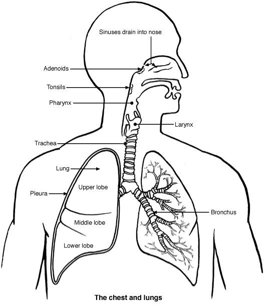
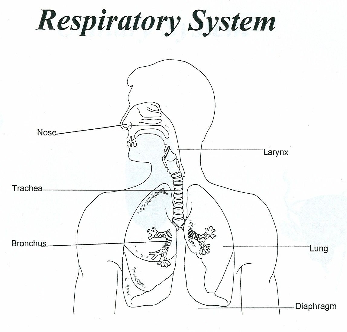
View Original Image at Full Size. Labeled diagram of the lungs/respiratory system. Image 37789 is a 1125 by 1408 pixel WebP Uploaded: Jan10 14. Last Modified: 2014-01-10 12:15:34

lungs diagram unlabelled Google Search Lunges, Arthritis, How to stay healthy
The right lung has 3 sections, called lobes. The left lung has 2 lobes. When you breathe: Air enters your body through your nose or mouth. Air then travels down the throat through the larynx and trachea. Air goes into the lungs through tubes called main-stem bronchi. One main-stem bronchus leads to the right lung and one to the left lung:
Respiratory System With Label Drawing at GetDrawings Free download
Anatomical Position and Relations The lungs lie either side of the mediastinum, within the thoracic cavity. Each lung is surrounded by a pleural cavity, which is formed by the visceral and parietal pleura. They are suspended from the mediastinum by the lung root - a collection of structures entering and leaving the lungs.

Lung Structure BioNinja
Root of lung. Lingula of left lung. Superior lobe (upper lobe) Middle lobe (only on right) Inferior lobe (lower lobe) Oblique fissure. Horizontal fissure of right lung. 144.30 Main bronchus. 152.23 Pulmonary ligament.
Respiratory System for Kids (Diagram + Theory + Vocabulary)
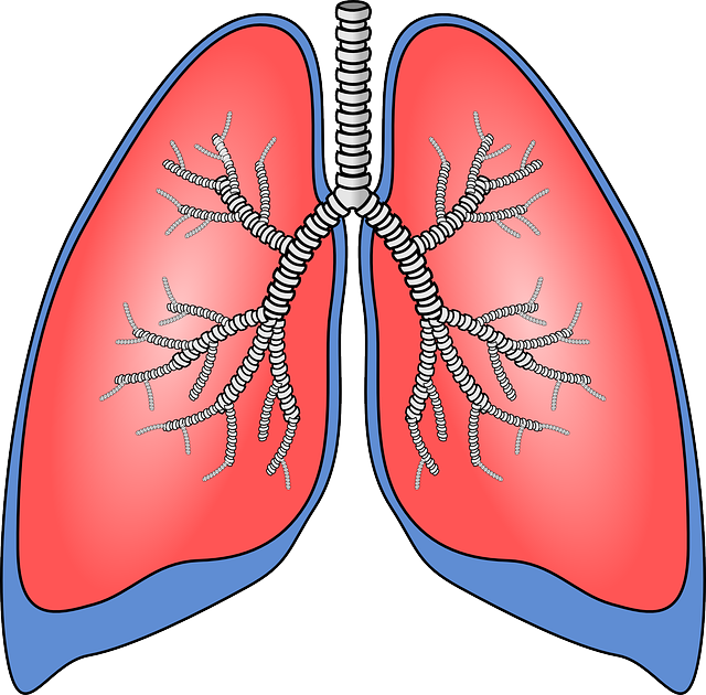
You inhale air into your mouth or nose. The air travels down the trachea (windpipe).; The air travels through the airways (bronchi) into your lungs.The air is directed through smaller and smaller passages (bronchioles).The air moves through a tiny duct (alveolar duct) and finally enters an individual alveolus (the singular of alveoli).; At this point, the oxygen molecules move through a single.
FileLungs diagram simple.svg Wikipedia
Respiratory system diagram The respiratory system How we breathe Respiratory conditions Summary The respiratory system allows air to reach the lungs, from which oxygen enters the blood.

Lungs Fill in the Blank Diagram Quizlet
The alveoli are located in the respiratory zone of the lungs, at the distal termination of the alveolar ducts. These air sacs are at the end points of the respiratory tract. There are approximately 700 million alveoli in the lungs, covering a total surface area of about 70 m 2, which is a considerably larger surface area relative to volume. The.

Download Unlabeled Lung Diagram Images Diagram Printabel
Diaphragm: The diaphragm is the main respiratory muscle that contracts and relaxes to allow air into the lungs. Last medically reviewed on July 31, 2023 How we reviewed this article: