Sinus Tarsi Syndrome Radsource
Sinus tarsi syndrome is the clinical syndrome of pain and tenderness of the lateral side of the hindfoot, between the ankle and the heel. Imaging often demonstrates the ligaments and soft tissues in the sinus tarsi are injured. Epidemiology

[PDF] Sonographically guided posterior subtalar joint injections via
Sinus tarsi syndrome is a clinical entity characterised by lateral hind-foot pain with worsening on palpation and weight-bearing, and perceived instability. It is associated with both traumatic and non-traumatic causes. Magnetic resonance imaging is the imaging modality of choice for assessment of the sinus tarsi and sinus tarsi syndrome.
Sinus Tarsi Syndrome Radsource
Abstract PURPOSE: To evaluate the tarsal sinus by using different imaging techniques and specialized planes. MATERIALS AND METHODS: Magnetic resonance (MR) imaging of the tarsal sinus was performed in 10 cadavers. Conventional arthrography of the anterior and posterior subtalar joints was then performed.
Sinus Tarsi Syndrome Radsource
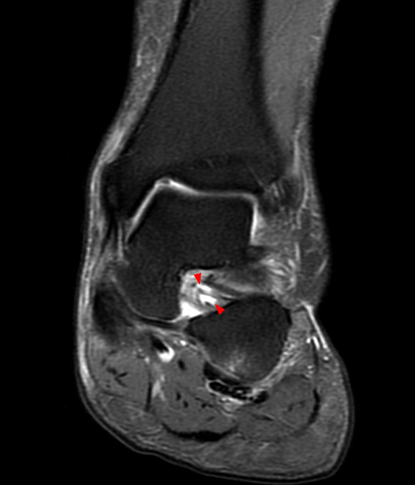
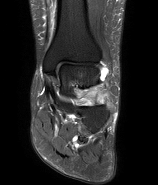
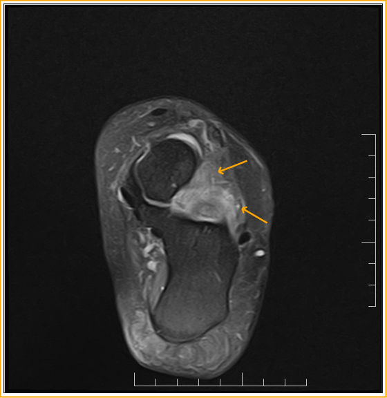
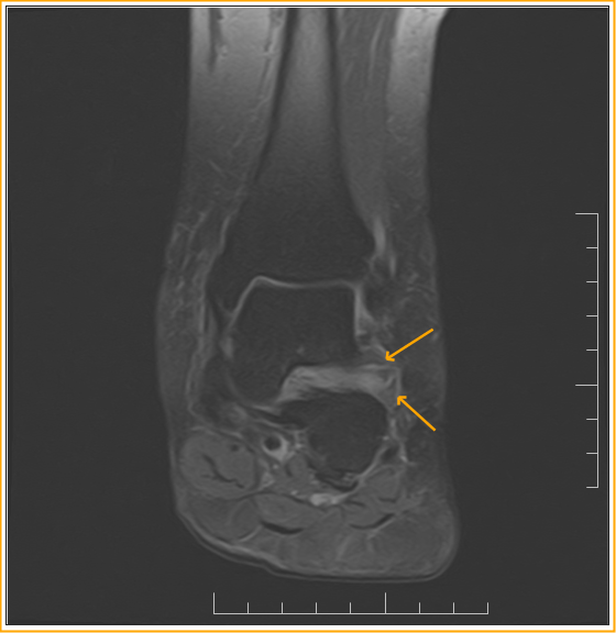
Age: 14 years Gender: Male mri Coronal T2 Coronal STIR Sagittal T1 Besides cystic lesions in the sinus tarsi, there is also talocalcaneal fibrous coalition with surrounding marrow edema. References 3 articles feature images from this case 16 public playlists include this case Related Radiopaedia articles Talocalcaneal coalition Tarsal sinus
Sinus Tarsi Syndrome Radsource
The Gruberi bursa, or the bursa mucosa of Gruberi, is a bursa located in the dorsal region of the ankle that extends from the sinus tarsi and along the frondiform ligament to surround extensor digitorum longus tendons. Terminology
Sinus Tarsi Syndrome Radsource
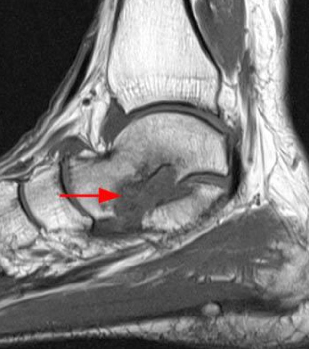
Abnormalities of the tarsal sinus and canal were seen on MR images in 33 cases (26.8%), were highly associated with tears of the lateral collateral ligament, and could be categorized according to the pathologic findings in patients with sinus tarsi syndrome: (a) diffuse infiltration with low T1- and T2-weighted signal intensity (n = 17) consiste.
Sinus Tarsi Syndrome MRI Sumer's Radiology Blog
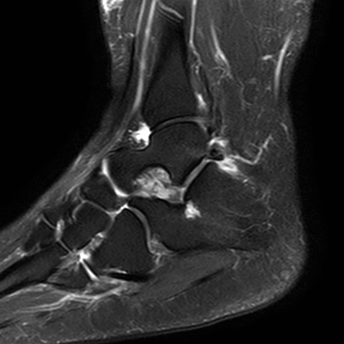
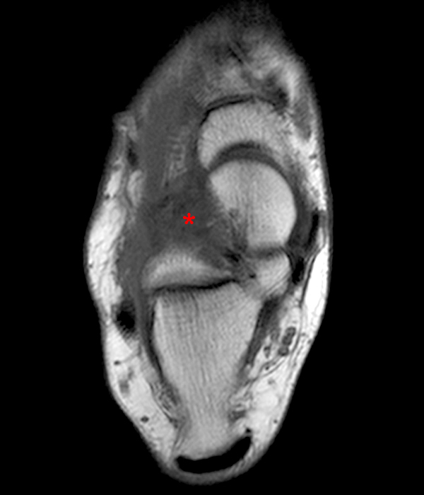
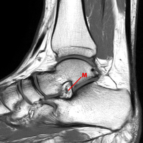
The sinus tarsi is a cone-shaped anatomical space bounded by the talus and calcaneus, continuing medially into the tarsal canal [1, 2].Along with fatty tissue, the tarsal sinus contains nerve endings, arterial anastomoses, posterior subtalar and talocalcaneonavicular joint recesses, joint capsules and five ligaments, i.e. the cervical ligament, the interosseous talocalcaneal ligament, and the.
Sinus Tarsi Syndrome MRI Sumer's Radiology Blog
Sinus tarsi syndrome (STS) is a clinical entity characterized by persistent anterolateral ankle pain secondary to traumatic injuries to the ankle. Historically, the etiology of this condition has not been well understood.
Sinus Tarsi Syndrome Radsource
Sinus tarsi syndrome is a clinical entity characterised by lateral hind-foot pain with worsening on palpation and weight-bearing, and perceived instability. It is associated with both traumatic and non-traumatic causes.. Clinical Radiology, Volume 76, Issue 12, 2021, pp. 940.e29-940.e35. A.W.H. Ng,., I.S.H. Ng.

Sinus tarsi syndrome Radiology Case
Sinus tarsi syndrome was first described in 1957 by Dr. Denis O'Connor [1]. The syndrome presents as pain in the lateral hindfoot, made worse following application of pressure to the opening of the sinus tarsi [2].. 2023, European Journal of Radiology. Show abstract. The sinus tarsi is a funnel-shaped region at the junction of mid-foot and.

Sinus Tarsi Syndrome Radsource
Sinus tarsi syndrome is used to describe a range of distinct underlying pathologies. MRI and arthrography show non-specific abnormalities in patients and identifying underlying pathologies is challenging. Discussion: The distinct range of underlying pathologies makes identifying specific imaging abnormalities and optimal treatments difficult.
Sinus Tarsi Syndrome Radsource
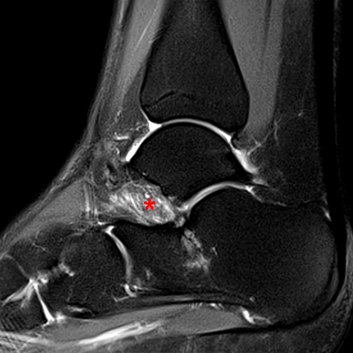
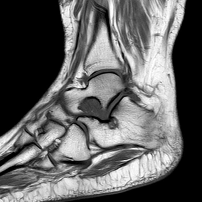
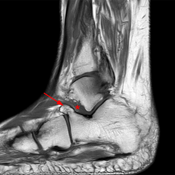
The sinus tarsi is a funnel-shaped region at the junction of mid-foot and hind-foot which contains fat, vessels, nerves and ligaments. The ligaments help stabilise the subtalar joint and maintain the longitudinal arch of the foot. The nerve endings contain proprioceptive fibres indicating a role for the sinus tarsi in movement of the foot.
Sinus Tarsi Syndrome Radsource
The sinus tarsi, also known as the tarsal sinus, is defined as the anatomical space between the neck of the talus and anterosuperior calcaneus. It is roughly cone-shaped, with the wider portion directed anteriorly and laterally.

Sinus Tarsi Pain Explanation The Foot and Ankle Centre of Victoria
Tarsal coalition is a congenital bridging of two or more tarsal bones. The bridging may be fibrous (syndesmosis), cartilaginous (synchondrosis), or osseous (synostosis).

Sinus Tarsi Syndrome MRI Sumer's Radiology Blog
The sinus tarsi (ST) is a funnel-shaped region at the junction of the mid-foot and hind-foot containing fat, vessels, nerves and ligaments. The ligaments function to stabilize the subtalar joint and help maintain the longitudinal arch of the foot.
MRI NEWSLETTER Sinus Tarsi Syndrome Radius Imaging
The sinus tarsi (ST) is a small. MR imaging of the tarsal sinus and canal: normal anatomy, pathologic findings, and features of the sinus tarsi syndrome. Radiology. 1993; 186 (1):233-240. doi: 10.1148/radiology.186.1.8416571. [Google Scholar] 2. Stoller DW, Ferkel RD. The ankle and foot. In: Stoller DW, editor.